

Thumb Ulnar Collateral Ligament Tear
Patient Education
Overview
Thumb Ulnar Collateral Ligament Tear is often associated with a sprained thumb that is caused by a fall onto an outstretched hand. The sudden force bends the thumb backwards causing the ligaments that support the thumb to stretch beyond their limits or tear. The ligament involves in most thumb sprains is called the ulnar collateral ligament (UCL) located on the inside of the knuckle joint. A tear to this ligament can be painful and may cause weakness and instability in the finger.
Treatment for thumb UCL tear is to keep the sprained thumb from moving for the ligament to heal with time. This is often done by wearing a splint or cast. However, more severe sprains may require surgery to repair the UCL and restore stability to the joint.
Description
The UCL is a strong, fibrous tissue that connects the metacarpal and the proximal phalange of the thumb at the metacarpophalangeal (MCP) joint. Sprains at this joint can range from a stretch (tiny tear) to a complete tear through the ligament.
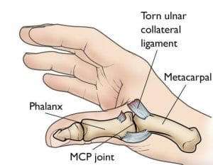
Fig. 1: Illustration of a completely torn ulnar collateral ligament.
Degrees of Thumb Sprains
Thumb sprains are graded based on the degree of injury to the ligaments:
- Grade 1 mild sprain: the ligaments are stretched but still intact.
- Grade 2 moderate sprain: the ligaments are partially torn and may accompany with some loss of function.
- Grade 3 severe sprain: the ligament is completely torn or is pulled off of the bone. This may cause an avulsion fracture, where the ligament pulls off a small piece of the bone. Significant injuries like such would require medical or surgical care.
Cause
Any strong force that bends the thumb backwards, away from the palm can cause the ULC to stretch or tear. It often caused by a fall onto an outstretch hand, commonly occurred in skiers when falling on the ski slopes with their hands strapped to a ski pole. It is also common in athletes who play sports that involve catching a ball, such as football, baseball, and basketball.
Symptoms
Level of pain may vary depending on the severity of the sprain. Thumb sprain may cause bruising, tenderness, and swelling around the base of the thumb. If the UCL is completely torn, the ruptured ligament may cause a lump inside the thumb. The thumb joint may feel loose and unstable, making it difficult to grasp objects between the thumb and index finger. If a thumb sprain does not improve after 48 hours, it is important to check with your doctor for proper diagnosis and treatment to prevent long-term complications, including chronic pain, instability, and arthritis.
Doctor Examination
Physical examination
Your doctor will ask you how and when the injury occurred as well as describing your symptoms. To determine the severity of the UCL tear, your doctor will gently move your thumb in different positions to test the stability of the MCP joint. If the joint is loose and unstable, it may indicate that the ligament is completely torn.
Imaging studies
X-rays: your doctor may order an x-rays of your thumb and hand to rule out an avulsion fracture or any broken bones.
Stress X-ray: during this x-ray imaging, your doctor will apply tension to the injured thumb to examine the stability of the MCP joint.
Other imaging types: a magnetic resonance imaging (MRI) scan or an ultrasound may be ordered to examine the severity of the injury on soft tissues.
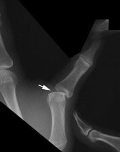 Fig. 2: Stress x-ray showing instability in the MCP joint, an indication that the ulnar collateral ligament is completely torn.
Treatment
Treatment for a sprained thumb varies with the severity of the injury.
Home Care
Mild thumb sprains can often be treated at home with the RICE protocol:
-
- Rest: avoid using your injured hand for at least 48 hours.
- Ice: you may apply a cold pack or a bag of ice wrapped in a paper towel immediately following the injury to reduce swelling. You may ice the affected area for 20 minutes at a time for several times a day.
- Compression: wear an elastic compression bandage to minimize swelling.
- Elevation: rest your hand above your chest as often as possible.
Nonsteroidal anti-inflammatory drugs (NSAIDs), such as aspirin or ibuprofen may also be used to reduce pain and swelling. See your doctor if symptoms are not improved after 48 hours.
Nonsurgical Treatment
Moderate thumb sprains can be treated at the doctor office by immobilizing the thumb joint. This can be accomplished with a bandage, thumb spica cast, or splint until the ligaments heal. Depending on its severity, you may be instructed to wear a splint or cast at all times.
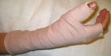 Fig. 3: Thumb spica cast on right arm.
Surgical Treatment
Severe thumb sprain may require surgery to restore stability to the MCP joint and help regain function. Thumb UCL may be repaired with InternalBrace Ligament Augmentation. This technique involves the reattachment of the ligaments to the bone using FiberWire. Surgery may also require repairing the avulsion fracture using a pin, screw, or bone anchor. Following the surgery, you may be required to wear a short arm cast or a splint for 6-12 weeks to keep the thumb immobilize while it heals.
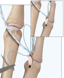
 Fig. 4: Thumb Ulnar Collateral Ligament repair with InternalBrace Ligament Augmentation.
Outcomes
When a sprained thumb is diagnosed and treated properly, it often recovers well without complications. However, being left untreated to heal on its own can lead to long lasting complications, including chronic instability, weakness, and arthritis. When this occurs, surgery is required to rebuild the ligament.
References:
Arthrex. (20180). Thumb UCL Repair with InternalBrace Ligament Augmentation: Surgical Technique. Retrieved from https://m.arthrex.com/resources/surgical-technique-guide/8IXwzkZmS0-ORwFWNzzj8w/thumb-ucl-repair-with-internalbrace-ligament-augmentation
Leversedge, F. & Jennings, C. (2018). Distal radius fractures: Broken wrist. OrthoInfo. Retried from https://orthoinfo.aaos.org/en/diseases–conditions/distal-radius-fractures-broken-wrist/
TR Johnson, LS Steinbach (eds.): Essentials of Musculoskeletal Imaging. Rosemont, IL. American Academy of Orthopaedic Surgeons, 2004, p. 378.